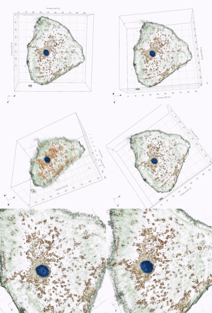
The International School of Frankfurt is one of the first schools in the world to incorporate the 3D Cell Explorer in their biology lessons. It is not a surprise that the students are much more engaged in their 3D biology lessons now then ever. Teachers have many new possibilities and students are working hands-on with this high end technology.
Rhiannon Wood, Upper School Principal and biology teacher at the International School of Frankfurt adds:
Biology is a naturally interesting subject for the many students who choose to study it, but with the arrival of our new 3D Cell Explorers student interest and engagement has moved to another level! Recently our grade 10 students carried out a cheek cell observation, which using a simple microscope in the past would have resulted in some mild interest as student could actually see (sometimes) one of their own cells. Using the 3D Cell Explorer students were able to observe bacteria on their cheek cell and measure the diameter, height and volume of the nucleus. A whole new world unfolded. Nanolive is allowing teachers to explore many new possibilities for investigations and IB biology students have access to a wider range of explorations for their IAs. Students love using high end tech in their school science lab and find it hugely exciting and motivating.

Animated cheek cell imaged with the 3D Cell Explorer
cell.academy is very excited to see that authentic 3D live cell images and videos in biology lessons inspire the scientists of tomorrow! Check the video below of how teachers are getting acquainted to the 3D CX after their arrival!
