Endocytosis: cell drinking and cell eating

Endocytosis: cell drinking and cell eating

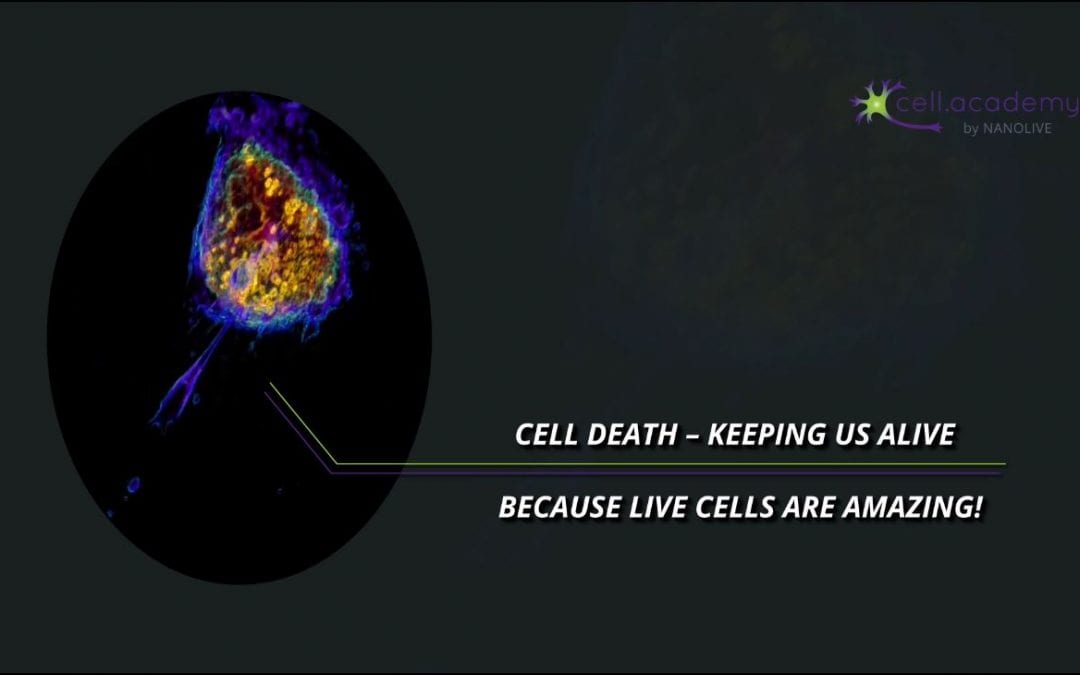
To introduce something into our body, we usually need to eat, drink, breathe or absorb through our skin. Did you know that this also happens at the cellular level? In this video, we will be talking about different forms of bulk transport into cells by illustrating these processes with videos of living cells, because live cells are amazing!

Eukaryotic cells are surrounded by a plasma membrane that defines the border between intracellular components and the extracellular environment. Plasma membranes are involved in many cellular aspects, but one of their main roles is to transport molecules, ions and other substances in and out of the cell.

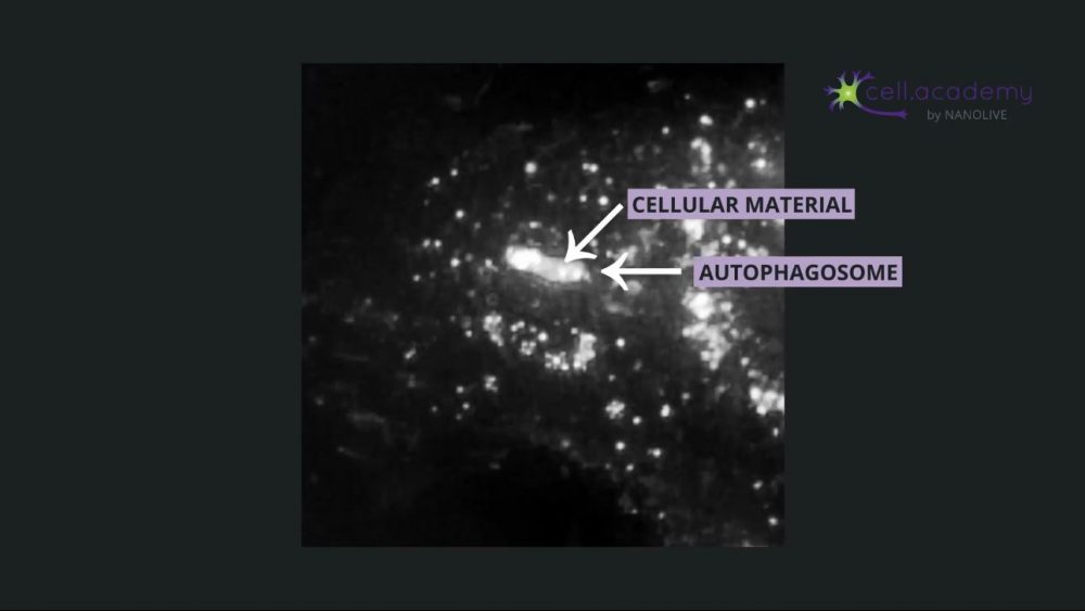
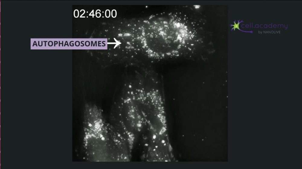
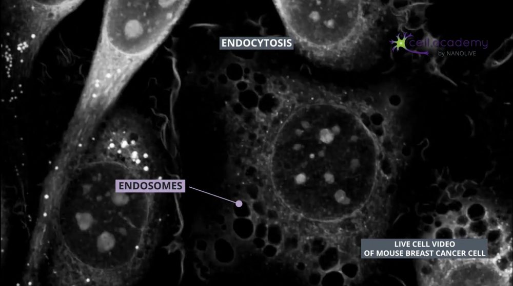
Endocytosis is a process in which membrane deformations lead to the generation of vesicles that can help uptake substances from the external environment.

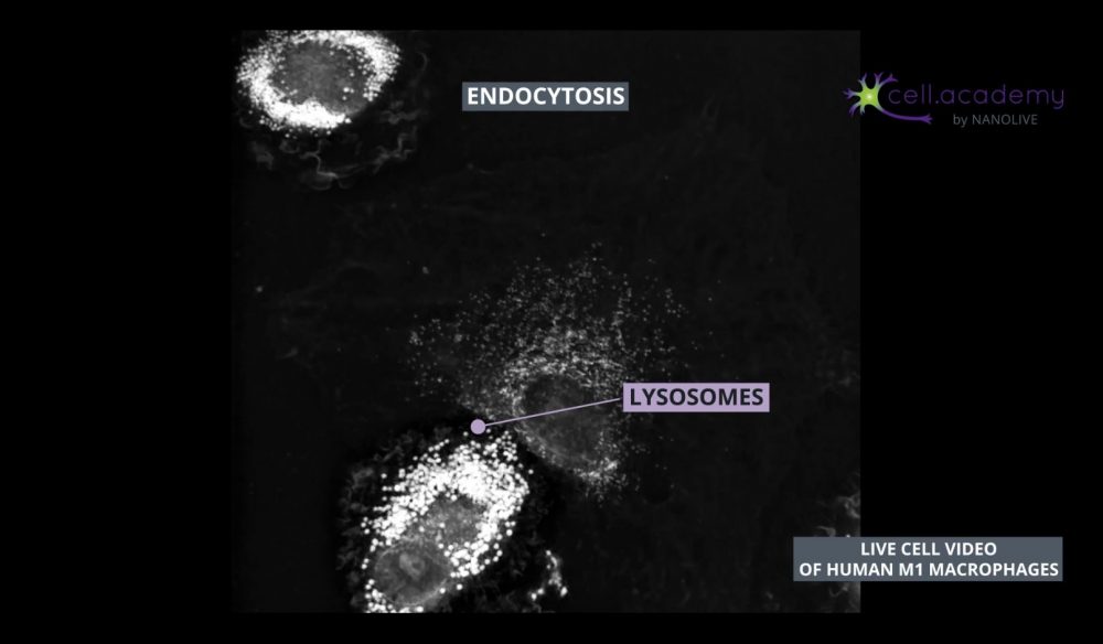
Endocytic vesicles fuse with early endosomes, which are structures used as transport vehicles inside the cell. These structures contain hydrogen pumps, which internalise hydrogen atoms from the cytoplasm, resulting in a gradual increase of endosomal acidity. Early endosomes can either be recycled back to the membrane or mature into late endosomes which can be found close to the Golgi apparatus, where they fuse with lysosomes. Finally, lysosomes are organelles that carry enzymes to degrade and help digest the uptaken material. Once digested, the content is further distributed to its final destination.

One specific form of endocytosis is called pinocytosis which is also known as “cell drinking”. This may give you a hint on what type of content is internalised in this process, right? As you may have guessed, during pinocytosis, the cell captures fluids like water and solutes. This is one way cells can internalise nutrients from the extracellular fluid.

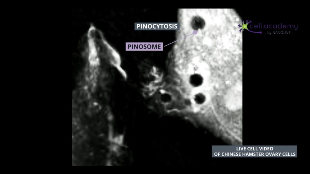
If we slow down this footage, you can observe how cytoskeletal reorganisation leads to the formation of plasma membrane ruffles. These ruffles fold, and form a pinosome: a vesicle which contains the fluid and solutes that the cell needs. Pinosomes will fuse with early endosomes, that will evolve into late endosomes and fuse in turn with lysosomes that will help them digest the content to be distributed, similar to the process described earlier.

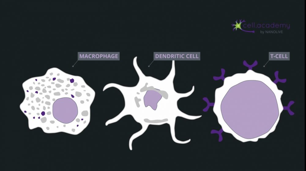
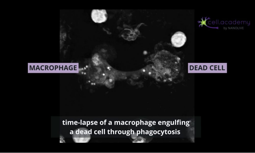
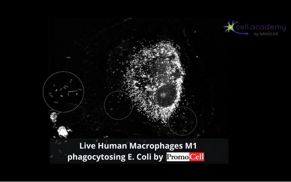
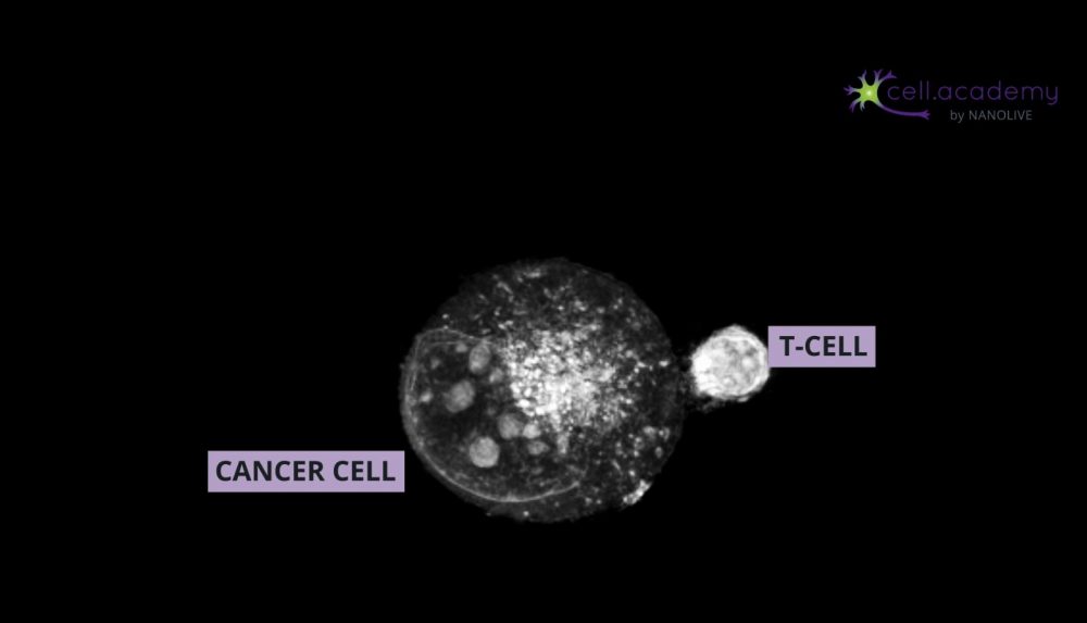
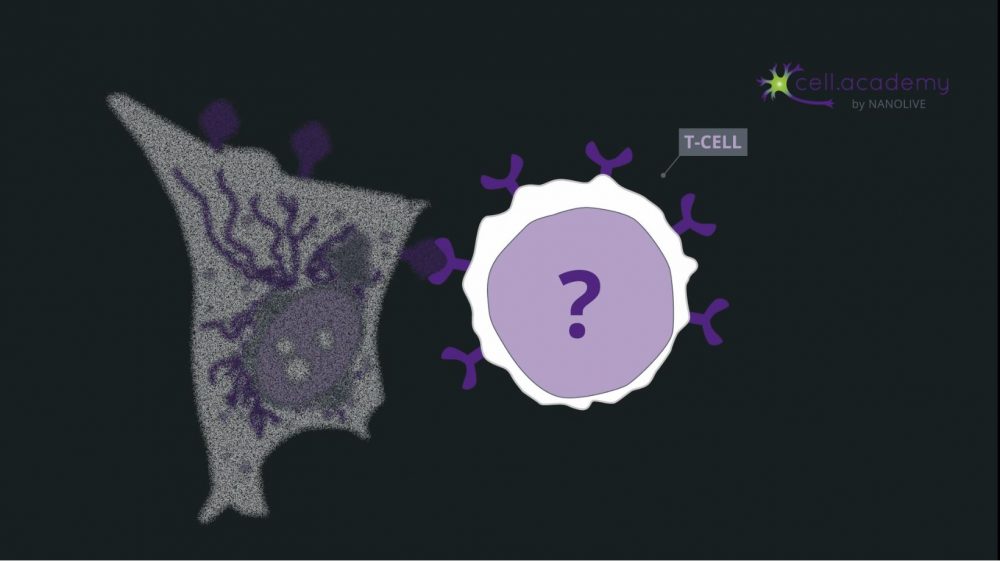
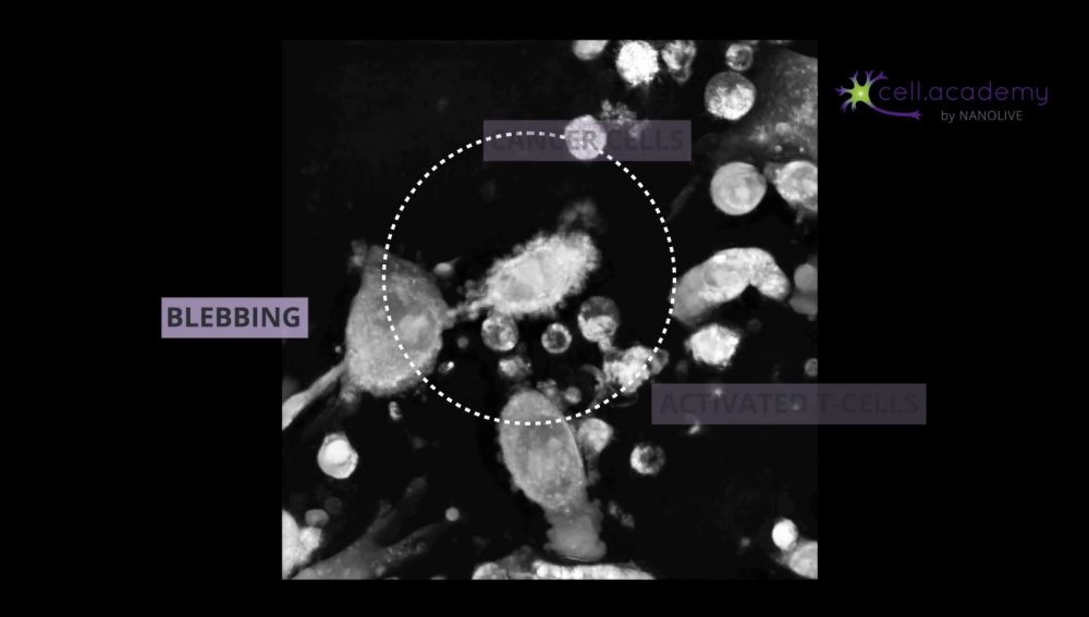
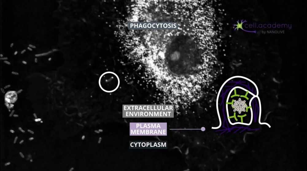
Larger particles such as microbes, bacteria and cellular debris are internalised in a process called phagocytosis, which you can consider as “cell eating”. While pinocytosis, is performed by almost all cellular types, phagocytosis is pretty much limited to immune cells, like macrophages.

During this process, particles are recognised by membrane receptors and internalised in structures called phagosomes. The content is later digested by enzymes provided in the fusion of phagosomes with lysosomes.  

So, as you have seen in this video, eating and drinking can also happen at the cellular level. Even though the internalised content is different, the general mechanism of endocytosis is preserved. Do you know of any other forms of endocytosis or bulk transport?

Blood related diseases

Blood related diseases

Did you know that around 7% of your body mass is blood? Blood plays a role in a wide variety of metabolic, homeostatic and immune processes. In fact, it is through blood that nutrients, hormones and waste are transported. In today’s video we will be talking about blood and its diseases, and we will illustrate our explanations with videos of living cells… because live cells are amazing!

The cellular components of blood are red blood cells, white blood cells and platelets. Platelets respond to blood vessel injury and help stop bleeding by creating a platelet plug. White blood cells, like the macrophages we showed in our previous video on the immune system (https://youtu.be/qmedkNV4NVY), are mainly involved in immune responses. And, finally, red blood cells help in distributing oxygen to the body tissues.

Anemia

Blood-related diseases are often due to small defects in these cells. If I asked you to list some common blood-related diseases, I am pretty sure that anemia would be one of the first to come up! Indeed, anemia is a common condition where problems in oxygen absorption result in fatigue and general weakness. Supplementing your diet with iron is generally enough to cure it, since iron plays a vital role in oxygen absorption. Seems easy, right? Well, there are some more complex types of anemia, like sickle cell anemia which is hereditary and harder to treat. Healthy red blood cells are round and flexible, making them perfect for oxygen transport. But, if you suffer from sickle cell anemia, your red blood cells acquire a sickle or crescent shape and become rigid. This shape is not ideal for oxygen absorption and, since this shape makes them less flexible, they may get stuck in small blood vessels and cause a clot. Besides, as it is an inherited condition, treatment must be followed for a lifetime.

Trypanosomiasis – the sleeping sickness

Anemia makes you feel tired, but this should not be confused with sleeping sickness, also known as trypanosomiasis, which is endemic in sub-Saharan Africa. Trypanosoma is a parasite which uses tsetse flies as a vector and is transmitted to humans through the bite of infected flies. When in the human bloodstream, trypanosoma replicates and targets red blood cells with the goal to manipulate and evade the immune system to allow survival and transmission. In this process, the parasite produces vesicles containing virulence factors that fuse with human red blood cells, causing membrane alterations, leading to anemia-like symptoms. In advanced stages, the disease attacks the central nervous system and, if not treated, can be fatal. 

Malaria

If you follow our channel regularly, this may sound familiar to you. In our Malaria Day video (https://youtu.be/bNE4qupNiq4), we explained that Malaria is caused by Plasmodium parasites that are transmitted to humans through the bite of infected female anopheles mosquitoes. Once in the bloodstream, Plasmodium also targets red blood cells, but with a different purpose than trypanosoma. Plasmodium evades the immune system by hiding within the red blood cells where they can proliferate undisturbed. Plasmodium feeds itself with the hemoglobin in host red blood cells, which eventually die. As you have probably understood by now, the loss of red blood cells leads to… extreme fatigue!

So, as we have seen today, fatigue can be explained by many factors, but it usually comes down to alterations in red blood cells. Do you know of any other blood-related diseases?

The Hallmarks of Cancer

The Hallmarks of Cancer

What is cancer? Cancer is when abnormal cells divide in an uncontrolled way. Sounds simple, right? Well, not really. Abnormalities are generally due to gene changes which impact the cell cycle. There are over 100 different types of cancer, so finding a unique cure is very difficult. However, in the year 2000, researchers Douglas Hanahan and Robert Weinberg published a ground-breaking paper with 6 properties that all cancerous cells have in common, which they called hallmarks. In this video, we will be explaining these 6 different hallmarks and illustrating them with videos of living cancer cells – because, live cells are amazing!

#1 Sustaining proliferative signaling

Let’s start with the first and most fundamental hallmark: Sustaining proliferative signaling. Cells have a receptor at their surface which, when receiving a growth factor signal from neighboring cells, initiates an intracellular cascade of signaling which leads to cell growth and division. Cells normally need this feedback from other cells to know when to divide, ensuring that proliferation happens in a coordinated manner.

Cancer cells are able to divide even without receiving these signals which leads to an uncontrolled proliferation of these abnormal cells. It is often a mutation in the receptor’s gene that maintains a growth signaling cascade – even in absence of the signal. At each replication cycle, the mutation is passed onto the daughter cells. This uncontrolled proliferation could eventually lead to the formation of a mass which we call a tumor.

#2 Evading growth suppressors

Which leads us to the second hallmark: evading growth suppressors. In order to maintain homeostasis, along with growth factors, cells can also produce growth suppressors which act as a stop signal for themselves or on neighboring cells if they are growing in an uncoordinated fashion. However, cancer cells, stubborn as they are, happen to ignore these “anti-growth signals” just like a speeding car going through a red light. This, also contributes to continuous proliferation of the cancerous cells.

#3 Resistance to cell death

So, these mutated cells are dividing without control, and as we saw in the previous video on cell death which you can watch on our channel, cells have an internal sensor to detect dangerous mutations, and initiate apoptosis to eliminate faulty cells through cell suicide. So you would think that these mutated cells would eventually die, right? Well that would be the ideal solution, however, the third hallmark of cancer is the resistance to cell death. Cancer cells often have a mutation in one of the signaling proteins leading to cell death, which helps them evade this fate.

#4 Enabling replicative immortality

If you remember, in the cell death video I had also mentioned that cells have a natural expiry date after which they are destined to die. But don’t get your hopes up, because it so happens that the fourth hallmark of cancer is enabling replicative immortality, in other words – some cancer cells overcome this expiry date and can divide indefinitely rendering them…. Immortal. Henrietta Lacks was a patient who died of cancer in 1951, and a cancer cell line called HeLa was derived from her cervical cancer cells and this line is still being widely used in research today due to its immortality.

#5 Inducing angiogenesis

Cell growth and division require a lot of energy which is provided in form of nutrients and oxygen from the surrounding blood vessels. So, you would think that at some point, cancer cells would not have enough resources to divide endlessly, especially if they are packed up in a tumor. Well, cancer cells secrete molecules which stimulate blood vessel growth. This process is called angiogenesis and is the fifth hallmark of cancer. This creates a larger network of vasculature around and within the tumor and provides the cells sufficient nutrients and oxygen to keep growing. Angiogenesis is a vital process in development that is usually activated during wound healing, but microenvironmental changes cause it to be activated in cancer cells as well.

#6 Activating invasion and metastasis

The sixth hallmark is also the event that most often leads to death: activating invasion and metastasis. At this stage, the cancerous cells not only invade neighboring tissues but also activate metastasis -which means these cells can now migrate through the blood and spread all around the body and eventually form new tumors in other locations. Therefore, it is difficult to locate them and provide focused treatment. I guess you’re starting to understand why it’s so complex to find a cure against cancer.

These six properties are all due to genetic modifications or changes in the microenvironment surrounding the cells that accumulate over time. It has now been 19 years since these hallmarks were established and even though they remain fundamental, more have been discovered since. Cancer is a vast field of research and new discoveries are made daily. Understanding these underlying mechanisms has already helped make great progress in the field of cancer therapy, as each hallmark is a potential target for treatment.

Do you know of any other hallmarks of cancer? Let me know in the comments below and head over to cell.academy to see the images of cancer cells we showed in this video…  and don’t forget to subscribe to our channel.


References:

Hanahan, D., & Weinberg, R. A. (2000). The Hallmarks of Cancer. Cell, 100(1), 57–70. https://doi.org/10.1016/s0092-8674(00)81683-9

Collins, K., Jacks, T., & Pavletich, N. P. (1997). The cell cycle and cancer. Proceedings of the National Academy of Sciences, 94(7), 2776–2778. https://doi.org/10.1073/pnas.94.7.2776

Cooper GM. The Cell: A Molecular Approach. 2nd edition. Sunderland (MA): Sinauer Associates; 2000. The Development and Causes of Cancer. Available from: https://www.ncbi.nlm.nih.gov/books/NBK9963/

Duesberg, P., & McCormack, A. (2013). Immortality of cancers. Cell Cycle, 12(5), 783–802. https://doi.org/10.4161/cc.23720

Pereira, B., & Ferreira, M. G. (2012). Sowing the seeds of cancer. Current Opinion in Oncology, 1. https://doi.org/10.1097/cco.0b013e32835b6358

Seyfried, T. N., & Huysentruyt, L. C. (2013). On the Origin of Cancer Metastasis. Critical Reviews™ in Oncogenesis, 18(1–2), 43–73. https://doi.org/10.1615/critrevoncog.v18.i1-2.40

Hainaut, P., & Plymoth, A. (2013). Targeting the hallmarks of cancer. Current Opinion in Oncology, 25(1), 50–51. https://doi.org/10.1097/cco.0b013e32835b651e

https://www.nature.com/scitable/topicpage/cell-division-and-cancer-14046590

MALARIA DAY 2019

MALARIA DAY 2019

Malaria is a life-threatening disease caused by Plasmodium parasites that are transmitted to people through the bites of infected female Anopheles mosquitoes.

While the species Plasmodium falciparum causes 99,7% of the cases in the African region, Plasmodium vivax accounts for 74,1% of estimated malaria cases in the Region of Americas. Nevertheless, malaria disease is less virulent and mortal when caused by P. vivax than when caused by P. falciparum. For this reason, even if there are around 14 different Plasmodium species causing disease in humans, malaria is frequently just linked to Plasmodium falciparum1.

But how is malaria transmitted through a mosquito if it is in fact Plasmodium the parasite causing it?

Parasites are organisms that live inside or on the surface of a host organism, which they use to help them survive.

Plasmodium falciparum infects female Anopheles mosquitoes with gametocytes (the precursors of female and male gametes or sexual cells) when one of them bites an individual suffering from malaria. Indeed, mosquitoes and humans are the two Plasmodium hosts.

Once at the mosquito’s intestine, P. falciparum undergoes multiplication (in a cycle known as the sporogonic cycle). As a result of this multiplication, structures known as sporozoites are formed and released. When biting an individual, the mosquito injects the sporozoites to the human bloodstream.

At that point, the human becomes its new host and two cycles take place: the first one in the liver (known as the exo-erythrocytic cycle), and the second one on the bloodstream (known as erythrocytic cycle).


In the liver, sporozoites infect liver cells, mature and form schizonts. Once schizonts burst, they release merozoites.

In the bloodstream, P. falciparum’s merozoites infect host red blood cells as a strategy to avoid being recognised by macrophages that would kill them2. They also digest host erythrocytes’ haemoglobin, as they need it to grow and to complete asexual replication. Under the microscope, haemoglobin loss in infected red blood cells is translated in a loss of brightness and density, compared to healthy erythrocytes3 (Figure 1).

Figure 1. Caption of live cell video showing malaria blood sample (infected red blood cell surrounded by healthy red blood cells)

During the erythrocytic cycle, merozoites mature into rings and from this stage and on, there are two possible options: either they evolve to form gametocytes, or they evolve and form new merozoites through intermediate stages (trophozoites and schizonts). Gametocytes will be again ingested by Anopheles mosquitoes, leading to a continuation of P. falciparum’s life cycle4,5.

Figure 2. Plasmodium falciparum’s erythrocytic cycle seen under the microscope. The different stages are highlighted and compared with traditional microscopy examples.

To know more about malaria’s erythrocytic cycle and how to diagnose malaria under the microscope click here! And if you want to refresh your knowledge about the immune system in general and macrophages in particular, do not hesitate to read our post and watch the video!

What’s the situation today?

At present, malaria causes millions of cases per year. 219 million cases were registered in 2017, which lead to 435 000 deaths, 266 000 of which were children under 5 years.

92% of the total cases were registered in the African region, which also accounted for 93% of the total deaths. Doubtlessly, although there are 87 countries around the world that have registered malaria, the African continent is the one suffering the strongest disease burden1.

Figure 3. 93% of total malaria cases are in Africa, where only 6 countries account for 55% of the total malaria cases registered in 2017.

What is the plan for the future?

The World Health Organization, whose main concern is international public health, developed a Global Technical Strategy for malaria, which aims to reduce the number of cases and deaths related to the illness by 90%, and increase malaria-free countries with at least 35 more countries, in a period from now until 2030.

In order to do so, vector control is one of the most important pillars. The use of insecticide-treated nets and indoor residual spraying, as well as drugs is crucial. This year, 3 pilot countries (Ghana, Kenya and Malawi) are introducing malaria vaccination1.

On this World Malaria Day, we want to raise awareness about the disease and its effects both in our cells and in a general perspective because, as this year’s key message reads, Zero malaria starts with me.


Bibliography:

  1. World Health Organization (https://www.who.int/campaigns/world-malaria-day/world-malaria-day-2019)Gomes, P. S., Bhardwaj, J., Rivera-Correa, J., Freire-De-Lima, C. G., & Morrot, A. (2016). Immune Escape Strategies of Malaria Parasites. Frontiers in microbiology7, 1617. doi:10.3389/fmicb.2016.01617
  2. Moore, L. R., Fujioka, H., Williams, P. S., Chalmers, J. J., Grimberg, B., Zimmerman, P. A., & Zborowski, M. (2006). Hemoglobin degradation in malaria-infected erythrocytes determined from live cell magnetophoresis. FASEB journal : official publication of the Federation of American Societies for Experimental Biology20(6), 747–749. doi:10.1096/fj.05-5122fje
  3. Bousema, T., & Drakeley, C. (2011). Epidemiology and infectivity of Plasmodium falciparum and Plasmodium vivax gametocytes in relation to malaria control and elimination. Clinical microbiology reviews24(2), 377–410. doi:10.1128/CMR.00051-10
  4. Shahinas, D.; Folefoc, A.; Pillai, D.R. Targeting Plasmodium falciparum Hsp90: Towards Reversing Antimalarial Resistance. Pathogens2013, 2, 33-54.
How cell death keeps us alive!

How cell death keeps us alive!

Why do cells die?

Cells in a general context, can die due to several factors. It could be due to external factors such as mechanical injury or exposure to toxins. Cells can also be attacked by the immune system if they are infected or faulty. Generally, given that billions of new cells are produced daily, it is important for older ones to die in order to maintain homeostasis which is a physiological balance.

How do cells die?

There are different ways a cell can die. In this blog post, we will be covering forms of both spontaneous and programmed cell death. If you want to see how pathogens and cancer cells are eliminated by the immune system, you can watch the dedicated video here: https://youtu.be/qmedkNV4NVY.

Necrosis – spontaneous cell death

Generally speaking, anything that modifies the pH levels in a cell can cause irreparable damage and provokes a spontaneous cell death called Necrosis. This can also be triggered by infection, toxins, injury or heat stress. Necrosis is considered as spontaneous, because it happens suddenly in response to trauma and is basically cell murder. 

Image 1

In image 1, a healthy mammalian cell is exposed to high concentrations of sodium hydroxide which increases the extracellular pH. This leads to osmotic perturbation which is lethal to the cell. You can see how the cell first gains in volume as the organelles swell up, which then causes a rupture of the plasma membrane, finally leading to an outflow of the intracellular contents. Sodium hydroxide is commonly found in drain cleaners, so make sure you take precautions when handling this powerful, but dangerous liquid!

Along with this outburst of contents, the cell also releases danger signals through molecules which activate an immune response to antigens in and around the dying cells shown in image 2.

Image 2

Necrosis is therefore essential to activate a rapid immune response following trauma. This response to injury is so stereotypical that pathologists often use this information to date the time of tissue injury, in the case of a heart attack, for example.

Apoptosis – programmed cell death

Many of our cells die at predetermined times through a programmed cell death called Apoptosis: in some cases, it’s because they are no longer needed after a certain stage of development. For example, in humans, during embryonic development, a webbing is present between our digits giving our hands and feet a “paddle” shape as seen in image 3.

Image 3

If you look at your hands now, this is generally not the case because this webbing is eliminated through apoptosis during the embryonic development. Now, you might know someone who has two fused fingers or toes, which is generally due to a lack of apoptosis during this stage and can only be corrected after birth through surgery.

In other cases, cells are intentionally produced in excess, in order to select the most adequate ones – a good example of this is during brain development: many neurons are produced but only those that make the correct connections with other neurons are preserved – the remaining are eliminated through apoptosis (see image 4).

Image 4

In general, most cells have a natural “expiration date” after which they are programmed to be eliminated and replaced by new ones. Red blood cells live for about four months, while skin cells only live about two or three weeks. Egg cells and most neurons are not replaced at all once they die… don’t hit anyone on their head – their brain cells might not recover!

So once a cell has reached the end of its predetermined life-time, it activates an internal pathway of apoptotic signals which leads to its own death – this could be thought of as cell suicide.

Image 5

In image 5, we can observe the morphological characteristics of apoptosis: the volume of the cell reduces as the cell internally condenses its structures and fragments the DNA and organelles. This “boiling” feature of the plasma membrane called blebbing is due to the breakdown of the cytoskeleton. Finally, the cell is broken down into apoptotic bodies that phagocytic cells can engulf and remove before the contents of the cell can spill out onto surrounding cells and cause damage to them. This is a very clean death that doesn’t require an immune response, which is why we don’t realize that it’s happening!

Apoptosis is very important in maintaining homeostasis since, as new cells are created, older ones need to be eliminated in order to maintain steady functions. In a tumor, it is common for the cancerous cells to block their own apoptotic pathways, which is why they keep growing and proliferating without control.

Autophagy – recycling damaged cells

When speaking about programmed cell death, the term autophagy often comes up.

Image 6

It is characterized by autophagosomes (see image 6) which are double-membraned vesicles containing cellular material to be degraded. Some dying cells do present autophagosomes, hence the association of this process with cell death, however autophagy itself is a fundamental process in the adaptation to starvation and an efficient survival strategy in times of physiological stress. So rather than committing suicide, the cell degrades and recycles its inner organelles and unused proteins to maintain homeostasis and metabolic functions and ensure its survival.

In image 7, you can see the vacuolization of the cytoplasm followed by heavy lysosomal activity within these autophagic cells. The large autophagosomes fuse with lysosomes, which degrade the organelles and proteins from the cell’s cytoplasm. Instead of killing off the entire cell, it is only replacing some cell parts.

Image 7

Nutrient deprivation is the key activator of autophagy, and it takes around 12 hours of fasting to initiate the process. Some studies show that cycles of fasting can reset and rejuvenate the human body, which is why fasting-diets are gaining in popularity. But it’s important to note that the scientific studies on the link between fasting and autophagy focus on its role in disease prevention and longevity, not weight loss, and this is still a field of intense research.

So, as we’ve seen today, it seems ironic, but our cells need to die in order to keep us healthy! Do you know about any other types of cell death? Let me know in the comments below, and head over to cell.academy to see more videos of living cells!


References:
(1) Kurien, B. T. (2004). Just a minute: incredible numbers at play at the macro and micro level. Canadian Medical Association Journal, 171(12), 1497–1497. https://doi.org/10.1503/cmaj.1040579

(2) Ding, D., Moskowitz, S. I., Li, R., Lee, S. B., Esteban, M., Tomaselli, K., … Bergold, P. J. (2000). Acidosis Induces Necrosis and Apoptosis of Cultured Hippocampal Neurons. Experimental Neurology, 162(1), 1–12. https://doi.org/10.1006/exnr.2000.7226

(3) Rock, K. L., & Kono, H. (2008). The Inflammatory Response to Cell Death. Annual Review of Pathology: Mechanisms of Disease, 3(1), 99–126. https://doi.org/10.1146/annurev.pathmechdis.3.121806.151456

(4) Malik, S. (2012). Syndactyly: phenotypes, genetics and current classification. European Journal of Human Genetics, 20(8), 817–824. https://doi.org/10.1038/ejhg.2012.14

(5) Suzanne, M., & Steller, H. (2013). Shaping organisms with apoptosis. Cell Death & Differentiation, 20(5), 669–675. https://doi.org/10.1038/cdd.2013.11

(6) Levine, B., Mizushima, N., & Virgin, H. W. (2011). Autophagy in immunity and inflammation. Nature, 469(7330), 323–335. https://doi.org/10.1038/nature09782

(7) Godar, R. J., Ma, X., Liu, H., Murphy, J. T., Weinheimer, C. J., Kovacs, A., … Diwan, A. (2015). Repetitive stimulation of autophagy-lysosome machinery by intermittent fasting preconditions the myocardium to ischemia-reperfusion injury. Autophagy, 11(9), 1537–1560. https://doi.org/10.1080/15548627.2015.1063768

(8) van Niekerk, G., du Toit, A., Loos, B., & Engelbrecht, A.-M. (2018). Nutrient excess and autophagic deficiency: explaining metabolic diseases in obesity. Metabolism, 82, 14–21. https://doi.org/10.1016/j.metabol.2017.12.007

(9) Mattson, M. P., Longo, V. D., & Harvie, M. (2017). Impact of intermittent fasting on health and disease processes. Ageing Research Reviews, 39, 46–58. https://doi.org/10.1016/j.arr.2016.10.005

Diving into the immune system!

Diving into the immune system!

The 2018 Nobel Prize for Physiology or Medicine went to James Allison and Tasuku Honjo for their research on cancer immunotherapy – to understand why their research was so revolutionary, we first need to go over some basics of the immune system and observe cells of the immune system in action, because live cells are amazing!

Macrophages, Dendritic Cells and T-cells

There are over 20 cell types in the immune system, each of which play multiple roles. In this video, we will be focusing on these three major cell types: Macrophages, Dendritic Cells and T-cells.

Macrophages – the cells that eat other cells

Macrophages, as their name suggests are large cells that eat others. Macrophages migrate through the bloodstream and tissues to detect pathogens or dead cells which they engulf through phagocytosis.

For example, if you cut yourself with a dirty knife, the bacteria will spread into your bloodstream where they encounter macrophages. In this caption below of a time-lapse of living cells, you can see E.coli bacteria being engulfed by a macrophage. Notice these shiny white dots – they are lysosomes, little organelles in the cell containing digestive enzymes which help digest the pathogen.

Dendritic cells – the antigen presenting cells

If the pathogen spreads and starts infecting our own cells, macrophages send an “alarm” through cytokines (chemical signals) to recruit their sister cells: dendritic cells.

Dendritic cells are also migrating, phagocytosing cells which serve as messengers to call in the heavy artillery. They are small and recognizable by elongated protrusions of their plasma membrane giving them a star shaped form. They engulf the pathogens through phagocytosis to process the pathogen’s antigen. Antigens are comparable to identity tags which are unique to each cell type. Dendritic cells expose the foreign antigen at their surface which they present to T-cells to activate them. Macrophages also have antigen presenting capabilities, so they complement the dendritic cells in T-cell activation.

T-cells – the killer cells

T-cells are small round cells containing a large nucleus. 

T-cells can also detect cancerous cells in the same way – you can see the close interaction between a T-cell and cancer cell here below.

Upon activation, T-cells duplicate and migrate to the site of battle to assist the macrophages. They can identify and kill the cells that have been infected by the pathogen thanks to the antigen they have been presented by the dendritic cells or the macrophages. 

The challenge of finding cures for diseases

So, if we have such a sophisticated system to fight against infections and invaders, why are researchers constantly working on finding cures for diseases? Well, many diseases which lead to serious health consequences are due to dysregulation of the immune system, which is why external intervention is required.

One example would be AIDS, Acquired Immune Deficiency Syndrome. It starts with a virus called HIV which attacks and gradually kills a subset of T-cells called CD4.

HIV treatment aims to increase the CD4 T-cell production, because if the number of CD4 T-Cells goes beneath a certain level, this can lead to AIDS, meaning part of the immune system is not functioning correctly and exposes the infected person to all kinds of infections and health issues.

Autoimmune diseases – a dysregulation of the immune system

Some diseases are not due to external factors, but from a dysregulation of our own immune system. These are called autoimmune diseases because the cells of our immune system attack our own healthy cells. One cause of this is often a defect in T-cells, which, upon encountering a cell which has a “self” antigen identity tag, misinterprets this antigen as foreign and eliminates it. This makes it difficult to find a cure for autoimmune diseases since anything that would reduce T-cell activity would also expose the body to infections that t-cells are supposed to fight.

Understanding the 2018 Nobel Prize in Physiology or Medicine

So, coming back to the 2018 Nobel Prize, now that we have a brief overview of some actors in the immune system. In some cases of cancer, a tumor develops because the immune system fails to recognize the mass of cancerous cells as foreign or dangerous due to an immune checkpoint on T-cells, which is a sort of brake system that interferes with the recognition.

The Nobel Prize laureates’ research consisted of discovering a drug which targets this checkpoint, resulting in a release of the break on these T-cells, so they can detect the cancer cells and kill them. The caption below shows how effective activated T-cells are in killing cancer cells. The blebbing is characteristic of a dying cell. 
With cancer being one of humanity’s greatest health challenges, this revolutionary research brings hope for the development of treatment.

Subscribe to our YouTube channel for more educational videos!